Muscle and adipose tissue segmentation in CT images |
|
Karteek Popuri, Howard Chung, Dana Cobzas, Jessica Lieffers, Agricultural, Food and Nutritional Science Martin Jagersand Vickie Baracos, Oncology |
ReferencesPopuri K., Cobzas D., Esfandiari N., Baracos B., Jägersand M. Body Composition Assessment in Axial CT Images using FEM-based Automatic Segment ation of Skeletal Muscle , Transaction of Medical Imaging ,2015 Popuri K., Cobzas D. and Jagersand M. A Fem Deformable Mesh for Active Region Segmentation, IEEE International Symposium on Biomedical Imaging (ISBI) 2013 Popuri K., Cobzas D., Jagersand M., Esfandiari N. and Baracos V. FEM-Based Automatic Segmentation of Muscle and Fat Tissues from Thoracic CT Images IEEE International Symposium on Biomedical Imaging (ISBI) 2013 Chung H., Cobzas D., Lieffers J., Birdsell L., and Baracos V. Automated segmentation of muscle and adipose tissue on CT images for human body composition analysis, SPIE Medical Imaging 2009 |
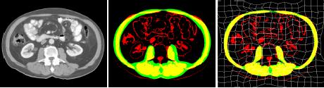
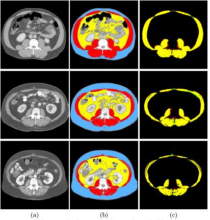
DescriptionThe ability to compute body composition in cancer patients lends itself to determining the specific clinical outcomes associated with fat and lean tissue stores. For example, a wasting syndrome of advanced disease associates with shortened survival. Moreover, certain tissue compartments represent sites for drug distribution and are likely determinants of chemotherapy efficacy and toxicity. CT images are abundant, but these cannot be fully exploited unless there exist practical and fast approaches for tissue quantification.We propose a fully automated method for segmenting muscle, visceral and subcutaneous adipose tissues, taking the approach of shape modeling for the analysis of skeletal muscle. Methodologically, we have two approaches, one based on a Finite Element Model and a Gaussian shape prior computing using PCA for encoding the muscle shape (TMI 2015, ISBI 2013) and an older approach based on a Free Form Deformation model (SPIE 2009). The shape model is learned from manually segmented images and used in conjunction with a tissue appearance prior.
|